輔助診斷臨床應用型 AI 及人工智慧模型自行建立平台
我們的解決方案,包含三大領域,並可相互整合:
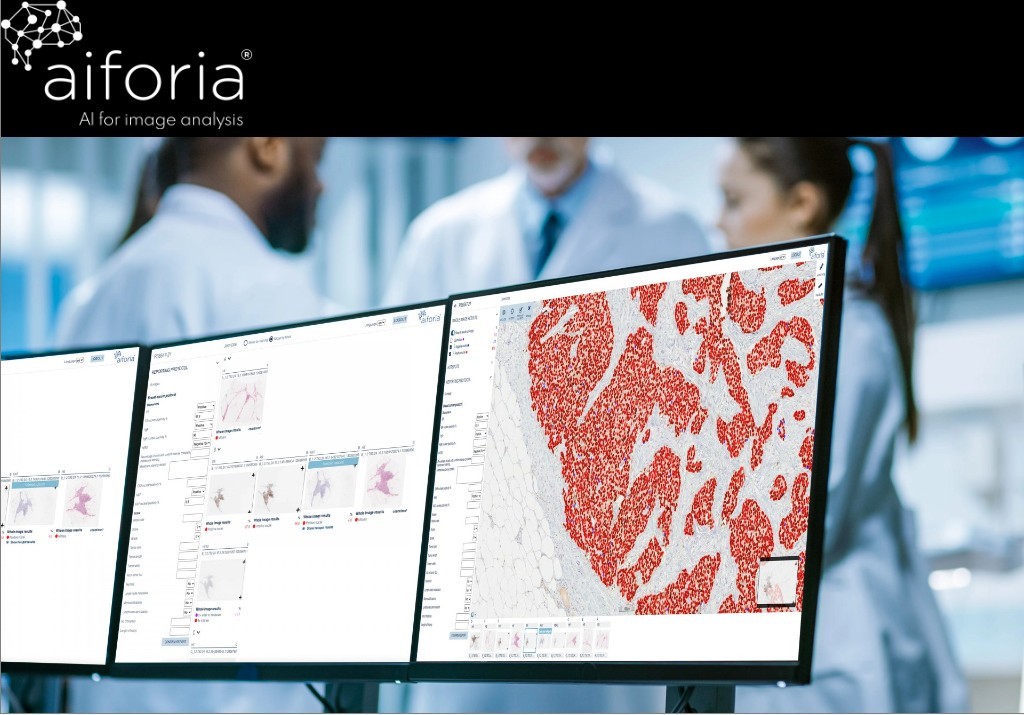
一、Aiforia Clinical Suite 人工智慧臨床診斷模組
•具備 CE-IVD 認證,無須重新進行訓練開發,可直接使用於臨床診斷。
•取得品質管理系統。
•Certified Quality Management System(ISO 13485)及資訊安全管理系統認證(ISO 27001)。
二、Aiforia Create 人工智慧自建模組平台
•僅需將玻片上傳至雲端平台,不需假他人之手,便可輕鬆建立自己的 AI 模型。
•利用此平台,我們可為您提供使用諮詢及教育訓練服務,後續的訓練及模型建立則可以由客戶自由進行,研究進度、概念及研究成果均掌握在客戶之手。
若您有意將您的模型轉化為商業模型,我們亦可提供此類協助。
三、Education Hub 雲端教學平台
•建立於雲端之教學平台,可提供玻片教育、閱片分享、雲端資料儲存及遠端會診功能。

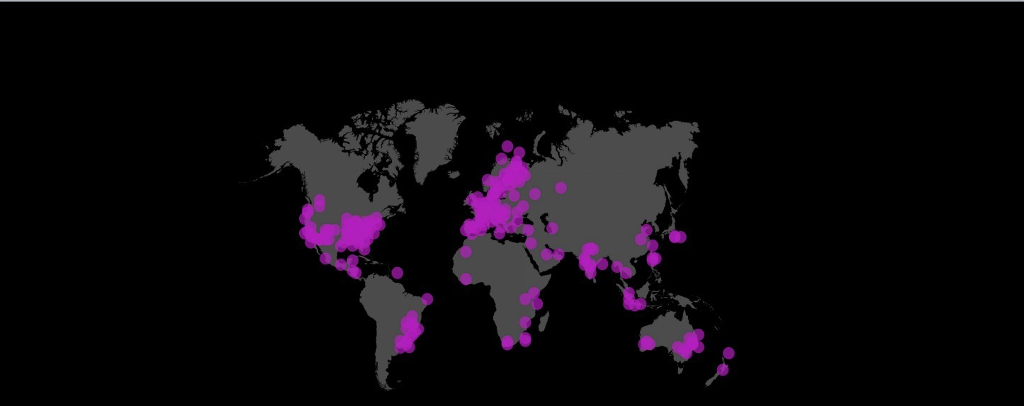

已有超過 2 百萬張影像使用 Aiforia 平台進行分析

全球超過 5,000 個用戶,其中包含全球的病理學家、醫師及研究人員使用 Aiforia 的 AI 及影像分析工具

已開發過超過 400 個以上用於影像分析的 AI 模型,超過 60 篇以上國際期合作發表刊

支援所有主要玻片掃描儀及顯微影像系統及 DICOM 格式,並可與 LIS、PACS 等平台進行整合
一、Aiforia Clinical Suite 人工智慧臨床診斷模組
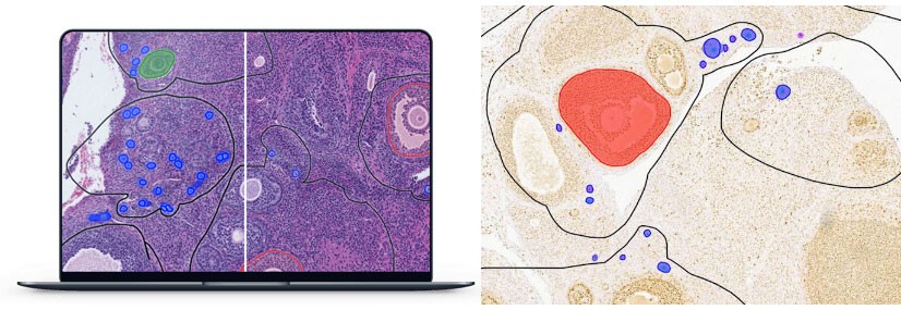
以 AI 工具輔助病理醫師提高診斷效率、降低瑣碎的量化分析工作、強化判讀精準度
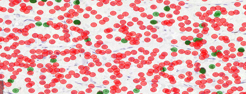
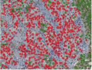
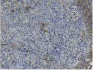
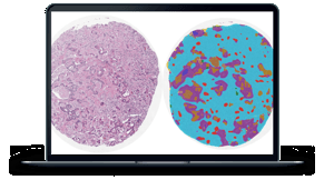
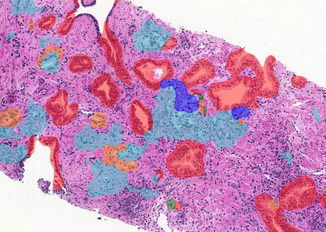
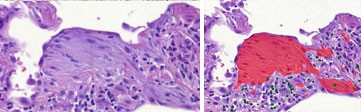
乳癌
乳癌
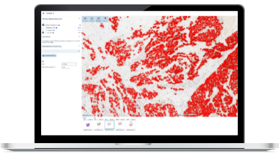
乳癌
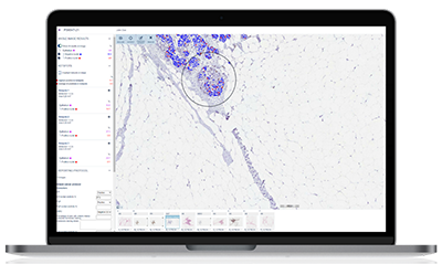
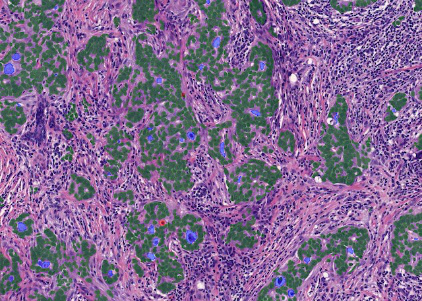
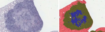
前列腺癌
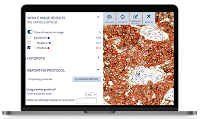
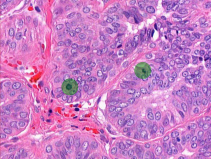
肺癌
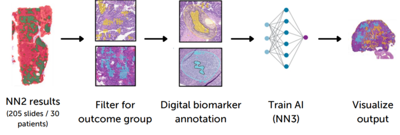
二、Aiforia Create人工智慧自建模組
AI 應用於研究領域
•可為任何 2D 影像(如病理組織切片、放射科影像)、各種影像格式(各家掃片機、顯微鏡影像系統、DICOM、他牌 AI 平台輸出影像格式)建立深度學習 AI 模型,進行定量分析。
•可為使用者量身打造 AI 模型 / 客制化服務。
•超過 60 篇以期刊論文,發表於神經科學領域、腫瘤 / 癌症領域及肝臟或其他醫學領域。
模型範例
Calculating Ki-67 proliferation index for pulmonary carcinoid tumors with AI at the Helsinki University Hospital.
Automated calculation and scoring of PD-L1 markers in a lung at Leiden University Medical Centre.
Creation of AI model to accurately classify high grade serous carcinoma into outcome groups at the University of Helsinki.
AI-assisted segmentation of colorectal carcinoma digitized images to identify prognostic and predictive histologic signatures at Mayo Clinic.
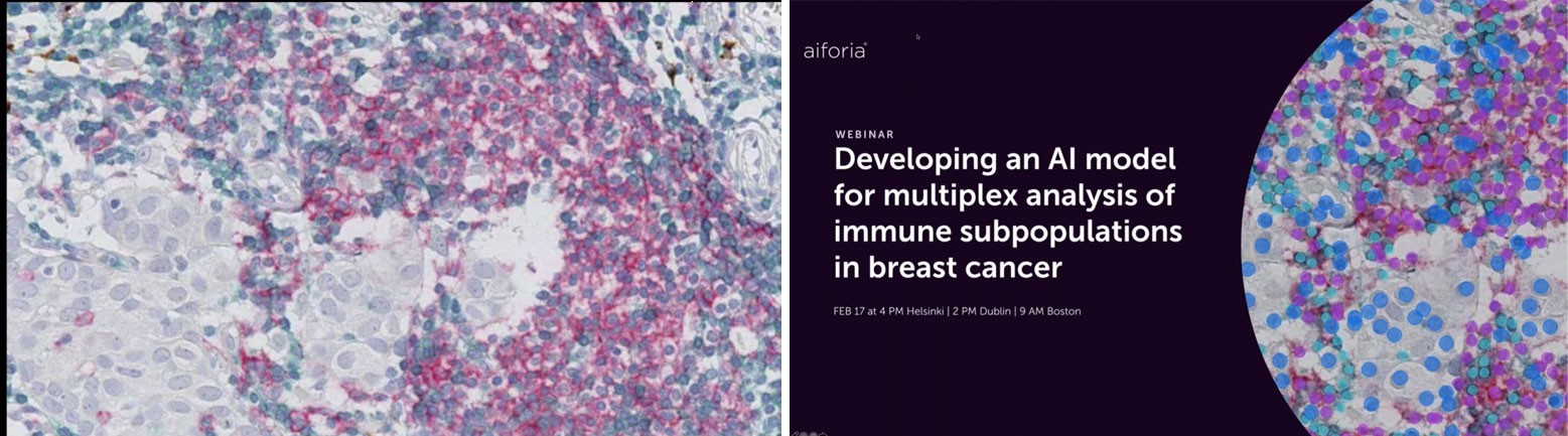
AI assisted detection of IHC stained CD3+ T-cells, CD20+ B-cells, DC-Lamp+ mature dendritic cells, and Hematoxylin in a HER2+ breast cancer surgical resection at the Royal College of Surgeons in Ireland.
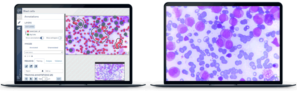
AI trained to identify blast cells and red blood cells on previously diagnosed cases of acute myelogenous leukemia at the Jinnah Sindh Medical University.
Automating tumor grading in non-small cell lung cancer(NSCLC)studies at MIT.
Automated tumor grading in prostate cancer biopsies at the Helsinki University Hospital, Finland, and in Region Skåne lab, Sweden.
Automated tumor grading in breast cancer biopsies.
Mitosis detection and quantification.
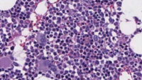
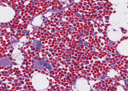
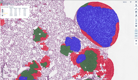
AI model used to identify and enumerate hematopoietic cells to determine changes in bone marrow cellularity for pharmacologic safety studies at Charles River Laboratories.
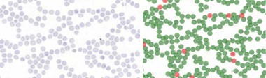
Blood Smear
Automated malaria parasite detection from blood smears at the University of Helsinki.
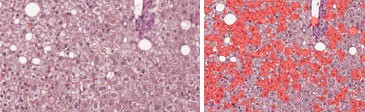
NAFLD and NASH
AI in studying nonalcoholic fatty liver disease(NAFLD)and its capability to segment structures in liver histology at the University of Helsinki.
NAFLD and NASH
Automated quantification of damage and scarring in liver tissue in nonalcoholic fatty liver disease(NAFLD)and nonalcoholic steatohepatitis(NASH).
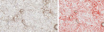
LUNG TISSUE
Identification and quantification of histopathological features of idiopathic pulmonary fibrosis(IPF)including fibroblast foci, and interstitial and alveolar inflammatory cells at the University of Helsinki.
LUNG TISSUE
Automated detection and quantification of cells and lesions in tuberculosis studies at Tufts University.
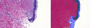
SKIN
Automated detection of dermis and epidermis of skin tissue sample.
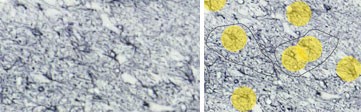
OVARIAN FOLLICLES
Preclinical studies on identification and quantification of the different stages of ovarian follicles in rats using AI models at Charles River Laboratories.
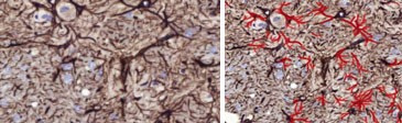
Quantifying astrocyte reactivity in preclinical neurotoxicity studies at Orion pharmaceutical company.
PARKINSON'S DISEASE
Use of AI for image analysis and astrocyte detection in Parkinson’s Disease research at Vall d'Hebron Instituto de Investigación.
ALZHEIMER’S DISEASE & CEREBRAL AMYLOID ANGIOPATHY
AI-assisted identification of the most characteristic markers of AD and CAA, including Aβ plaques, at Massachusetts General Hospital.
PARKINSON'S DISEASE
Quantitative assessment of alpha-synuclein pathology in preclinical Parkinson's disease studies at Lundbeck pharmaceutical company.
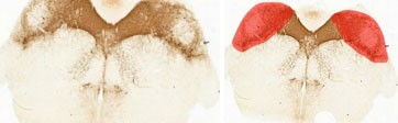
Automated nigra area detection.
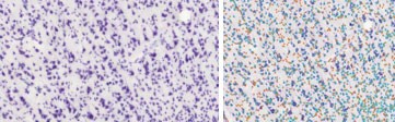
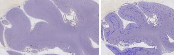
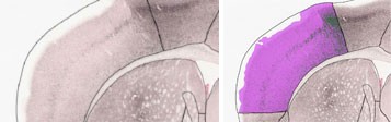
Grey/White matter segmentation and Neuron/Glia counts in human brain sample.
PARKINSON’S DISEASE
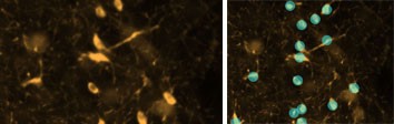
Automated dopaminergic neuron quantification in Parkinson’s disease research at Duke University.
三、Education Hub 雲端教學平台
•此平台是專為組織學、解剖學、病理學、寄生蟲學及獸醫病理學等課程所設計的。
•可以使老師及學生均不再受限於顯微鏡、固定的教室及書面資料。
•雲端平台可以接受老師使用任何玻片掃描儀所上傳的任何影像,並直接與學生分享。
•老師可以於平台中加入影像的註解、說明及影像的病史、個案背景訊息等,完整整個樣本的起源及狀態描述,豐富學生的學習資源。
•此平台基於軟體即服務 SaaS(Software-as-a-Service)模式,可以由簡單的研討會模式擴展延伸至大型進階版的教學模式。